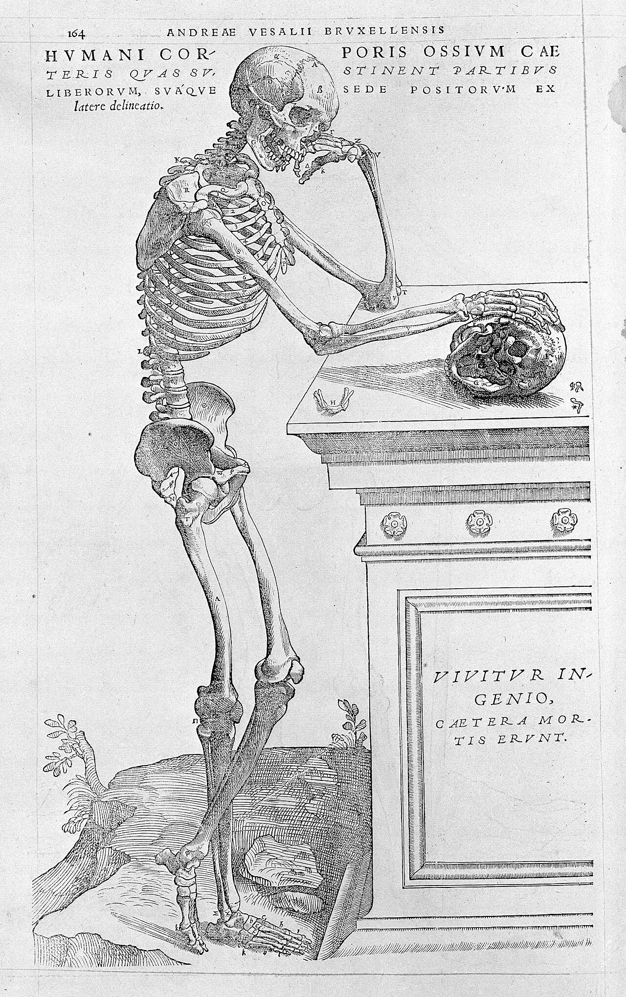

A baby has more bones than an adult.
The number of bones in an adult is not a fact of nature: it is a convention.
This time, the box with blurred edges lies within the human body.
Bones that fuse
A newborn has markedly more bones than an adult.
Not that it loses any: several fuse during growth. Separate pieces, which allow the body to grow and change shape, end up merging into a single one.
The skull, for example, is not all of a piece at birth.
And the number for an adult?
School generally teaches a precise figure.
This figure is a convention, for the following reasons.
It depends on what one decides to count as a bone. Should two pieces that are fused but still visible be counted separately? What about the small bones that form in certain tendons, and that not everyone has? What about the variations from one person to another — for there are some, and they are normal?
Depending on the answers, the total changes.
How many bones does a skeleton have?
The usual answer is 206.1
This number is a textbook value, not a count carried out on a person. Depending on the person, the actual count can differ from it by a few bones.
Why the number varies
- Sesamoid bones. These are small bones that form inside certain tendons, partly as a result of mechanical stresses. In other words, their number may depend in part on the strains the body undergoes.
- Wormian bones. Small extra bones that sometimes appear between the plates of the skull. This is most often a normal variation, with nothing pathological about it.
- Vertebrae. A non-negligible proportion of people have 23 or 25 vertebrae above the sacrum, instead of the expected 24.2
The mise en abyme is striking: the percentage itself varies with the way of counting. A study of more than three thousand whole-body radiographs finds 8.8% — of whom 5.5% with twenty-five vertebrae, 3.3% with twenty-three, and a single person with twenty-six. Other work measures something else, “transitional” vertebrae: a 2010 review reports, depending on the study, from 4 to 30%.3
Where does such a gap come from? First of all, because the same thing is not being counted: here segments above the sacrum, there transitional vertebrae, classified according to their own criteria.4
The figure that measures variation is itself variable. One would be hard pressed to find a better demonstration of the point of this page.
Why it really matters
This detail might seem inconsequential. Yet it has a consequence, and a serious one.
A surgeon who has to operate on a specific vertebra locates it by counting. If they count from the bottom and the person has one vertebra more or one fewer, they may operate at the wrong level.5
This is why specialists recommend, in such cases, imaging of the entire spine, rather than a partial count that can easily mislead.6
This is what a conventional count can cost when it is applied without checking who it is being applied to.
And at birth?
A newborn has between 275 and 300 bones.7
It loses none of them: some fuse as it grows. A baby’s skull is made up of several movable pieces, which allows it to pass through at birth and then lets the brain grow. The sacrum, for its part, sometimes completes its fusion only at around thirty.8
The skeleton is therefore not complete until an age at which one has long considered oneself an adult.
What this figure teaches
206 is not wrong. It is a reference value, useful for teaching — not an exact description of anyone.
Many common numbers are of this kind. They describe a typical case, and the typical case exists in no one in particular.
There is nothing abnormal about not having 206 bones. It is the figure that is a simplification, not the skeleton that is abnormal.
Bone is alive
Another received idea deserves to be corrected, and this one changes everything.
A museum skeleton looks like a framework: dry, hard, inert. It is tempting to conclude that bones are dead parts that hold up the rest.
A bone is a living tissue. Blood vessels run through it. It is constantly broken down and rebuilt, throughout life.
The essential point is this: what one sees in a museum is not what a living body contains. It is what is left of it when everything that was alive has gone.
What a skeleton is for
It fulfils several functions at once, of which generally only one is remembered.
It supports: without it, a human body would collapse like a garment dropped on the floor. It protects: the skull encloses the brain, the rib cage the heart and lungs, the spinal column the spinal cord. It enables movement, not by itself — a bone does not move on its own — but by serving as a lever for the muscles attached to it. It serves as a store of minerals, including calcium and phosphorus. It makes blood: inside the bones, especially the flat bones and the vertebrae in adults, the bone marrow produces more than two million red blood cells per second, without ever stopping. This is indispensable: a red blood cell lives no more than a hundred and twenty days.9
This last function is the one most often forgotten, and it is the most surprising: the body’s framework is also its blood factory.
The joints
A completely fused skeleton would be useless. What makes movement possible are the joints: the places where two bones meet.
They do not all move in the same way: their shape determines the movements they allow.
- The ball-and-socket joint, such as the shoulder or the hip: a ball in a cavity. It is the freest joint — the arm can rotate in almost every direction.
- The hinge, such as the elbow or the knee: it bends, essentially, in only one plane, like a door. Forcing it sideways is a common cause of sports injuries.
- The pivot, as between the first two vertebrae of the neck: it allows the head to turn from left to right.
- Fixed joints, such as those of the skull. They exist, but do not move once growth is over — these are the sutures between the bones of the skull, mentioned at the beginning of this page.10
At the points of contact, the ends of the bones are covered with cartilage, a smooth, supple tissue that absorbs shocks and lets the surfaces slide over each other. A thick fluid, synovial fluid, completes the arrangement — it acts as a lubricant. Around them, ligaments connect the bones to each other and prevent the joint from going too far.
Cartilage barely repairs itself. Unlike bone, which rebuilds itself throughout life, it contains no blood vessels: its cells are nourished by simple diffusion through the material that surrounds them. This is slow, and it is enough for maintenance but not for repair. Once worn, it does not come back. This is one of the reasons why osteoarthritis becomes more common with age.11
Precisely: what bones have taught us
This leads to an observation that this site comes across everywhere.
Bones are preserved. Soft tissues, almost never.
Everything we know about ancient bodies — human or animal — comes through what the bones have chosen to keep. It is not what existed, but what survived.
Yet this has made considerable discoveries possible. It was by reading bones that a fact recounted on our page about agriculture was established: in many regions, the skeletons of the first farmers are smaller and less well nourished than those of the hunter-gatherers who preceded them.12
Not everywhere, but in many regions — and that was enough to prompt a revision of a whole story that had been thought simple.
Bones speak, to those who know how to read them.
A rule that applies to this page
This page describes bodies.
Yet bodies vary — far more than anatomical plates suggest, with their single, perfectly symmetrical skeleton.
Some people have extra bones, others fewer. Some live with a bone disease, a prosthesis, a wheelchair.
This site will never write that there is a normal body from which the others deviate. Variation is the fact. The average is a calculation.
Note 1
For adults, there are 206 bones in the skeleton.
Betts, J. Gordon et al., Anatomy and Physiology, OpenStax, Rice University
Note 2
Sesamoid bones of the hand are variable but are found constantly at the metacarpophalangeal articulation of the thumb. […] Sutural bones are usually small, irregularly shaped ossicles, often found in the sutures of the cranium […] As many as 172 sutural or wormian bones have been found in one skull.
Bergman, Ronald A., Afifi, Adel K. and Miyauchi, Ryosuke, Illustrated Encyclopedia of Human Anatomic Variation, University of Iowa
Transitional anatomy, defined as having fewer or more than 24 presacral segments, was observed in 279 patients (8.8%).
Sabri, Shahbaaz A. et al., “Implications of Lumbosacral Transitional Anatomy on Vertebral Numbering: Evaluation of 3147 Adult Full-Length Spine Radiographs”, Neurosurgical Focus, vol. 58, no. 6, 2025, article E13
Note 3
Specifically, 174 patients (5.5%) had 25 presacral segments, 104 patients (3.3%) had 23, and 1 patient (0.03%) had 26.
Sabri, Shahbaaz A. et al., “Implications of Lumbosacral Transitional Anatomy on Vertebral Numbering: Evaluation of 3147 Adult Full-Length Spine Radiographs”, Neurosurgical Focus, vol. 58, no. 6, 2025, article E13
LSTVs are common in the general population, with a reported prevalence of 4%–30%
Konin, Gabrielle P. and Walz, Daniel M., “Lumbosacral Transitional Vertebrae: Classification, Imaging Findings, and Clinical Relevance”, American Journal of Neuroradiology, vol. 31, 2010, p. 1778-1786
Note 4
In 1984, Castellvi et al […] described a radiographic classification system identifying 4 types of LSTVs on the basis of morphologic characteristics (Fig 2). […] Although useful for characterizing the relationship between the transitional segment and the level above or below, this classification system does not provide information relevant to accurate enumeration of the involved segment.
Konin, Gabrielle P. and Walz, Daniel M., “Lumbosacral Transitional Vertebrae: Classification, Imaging Findings, and Clinical Relevance”, American Journal of Neuroradiology, vol. 31, 2010, p. 1778-1786
Transitional anatomy, defined as having fewer or more than 24 presacral segments
Sabri, Shahbaaz A. et al., “Implications of Lumbosacral Transitional Anatomy on Vertebral Numbering: Evaluation of 3147 Adult Full-Length Spine Radiographs”, Neurosurgical Focus, vol. 58, no. 6, 2025, article E13
Note 5
Errors in vertebral numbering can have devastating implications for patients, especially when intraoperative numbering errors occur.
Sabri, Shahbaaz A. et al., “Implications of Lumbosacral Transitional Anatomy on Vertebral Numbering: Evaluation of 3147 Adult Full-Length Spine Radiographs”, Neurosurgical Focus, vol. 58, no. 6, 2025, article E13
Note 6
complete spinal imaging is essential for accurate vertebral numbering, as partial counts based on ribs or fluoroscopic references can easily lead to errors
Sabri, Shahbaaz A. et al., “Implications of Lumbosacral Transitional Anatomy on Vertebral Numbering: Evaluation of 3147 Adult Full-Length Spine Radiographs”, Neurosurgical Focus, vol. 58, no. 6, 2025, article E13
Note 7
A newborn has 275 to 300 bones, while most adults have 206.
Cleveland Clinic, “How Many Bones Do Babies Have?”, 8 February 2024
A newborn has 275 to 300 bones, while most adults have 206.
Cleveland Clinic, “How Many Bones Do Babies Have?”, 8 February 2024
Note 8
Sacral fusion begins around puberty and is completed by age 25 to 30.
Sattar, Mohammad H. and Guthrie, Stuart T., “Anatomy, Back, Sacral Vertebrae”, in StatPearls, StatPearls Publishing, updated 30 July 2023
Note 9
The skeletal system is the body system composed of bones and cartilage and performs the following critical functions for the human body: • supports the body • facilitates movement • protects internal organs • produces blood cells • stores and releases minerals and fat
Betts, J. Gordon et al., Anatomy and Physiology, OpenStax, Rice University
Production of erythrocytes in the marrow occurs at the staggering rate of more than 2 million cells per second.
Betts, J. Gordon et al., Anatomy and Physiology, OpenStax, Rice University
Erythrocytes live up to 120 days in the circulation
Betts, J. Gordon et al., Anatomy and Physiology, OpenStax, Rice University
Note 10
At a pivot joint, a rounded portion of a bone is enclosed within a ring […] In a hinge joint, the convex end of one bone articulates with the concave end of the adjoining bone […] The joint with the greatest range of motion is the ball-and-socket joint. […] An immobile or nearly immobile joint is called a synarthrosis.
Betts, J. Gordon et al., Anatomy and Physiology, OpenStax, Rice University
Note 11
Cartilaginous tissue is avascular, thus all nutrients need to diffuse through the matrix to reach the chondrocytes. This is a factor contributing to the very slow healing of cartilaginous tissues.
Betts, J. Gordon et al., Anatomy and Physiology, OpenStax, Rice University
Note 12
entrée 2.9
entry 2.9
The fact concerning the skeletons of the first farmers, less well nourished than the hunter-gatherers who preceded them, is established in entry 2.9, with its sources
Notes and sources
- ↑
Passage The usual answer is 206.
For adults, there are 206 bones in the skeleton.
Source Betts, J. Gordon et al., Anatomy and Physiology, OpenStax, Rice University, chapter 7.1, “Divisions of the Skeletal System”, 1st paragraph (2nd ed.).
Open access open the source · checked on 27 October 2026
- ↑
Passage Sesamoid bones […] Wormian bones. Small extra bones that sometimes appear between the plates of the skull.
Sesamoid bones of the hand are variable but are found constantly at the metacarpophalangeal articulation of the thumb. […] Sutural bones are usually small, irregularly shaped ossicles, often found in the sutures of the cranium […] As many as 172 sutural or wormian bones have been found in one skull.
Source Bergman, Ronald A., Afifi, Adel K. and Miyauchi, Ryosuke, Illustrated Encyclopedia of Human Anatomic Variation, University of Iowa, Opus V (Skeletal System), entry “Wrist and Hand: Carpals, Metacarpals and Phalanges”, paragraph on the sesamoid bones of the hand; entry “Occipital Bone” (Cranium), paragraph beginning “Sutural bones are usually small”.
Open access open the source · checked on 27 October 2026
Passage A non-negligible proportion of people have 23 or 25 vertebrae above the sacrum, instead of the expected 24.
Transitional anatomy, defined as having fewer or more than 24 presacral segments, was observed in 279 patients (8.8%).
Source Sabri, Shahbaaz A. et al., “Implications of Lumbosacral Transitional Anatomy on Vertebral Numbering: Evaluation of 3147 Adult Full-Length Spine Radiographs”, Neurosurgical Focus, vol. 58, no. 6, 2025, article E13, abstract.
Open access open the source · checked on 24 October 2026
- ↑
Passage 8.8% — of whom 5.5% with twenty-five vertebrae, 3.3% with twenty-three, and a single person with twenty-six
Specifically, 174 patients (5.5%) had 25 presacral segments, 104 patients (3.3%) had 23, and 1 patient (0.03%) had 26.
Source Sabri, Shahbaaz A. et al., “Implications of Lumbosacral Transitional Anatomy on Vertebral Numbering: Evaluation of 3147 Adult Full-Length Spine Radiographs”, Neurosurgical Focus, vol. 58, no. 6, 2025, article E13, abstract.
Open access open the source · checked on 24 October 2026
Passage Other work measures something else, “transitional” vertebrae: a 2010 review reports, depending on the study, from 4 to 30%.
LSTVs are common in the general population, with a reported prevalence of 4%–30%
Source Konin, Gabrielle P. and Walz, Daniel M., “Lumbosacral Transitional Vertebrae: Classification, Imaging Findings, and Clinical Relevance”, American Journal of Neuroradiology, vol. 31, 2010, p. 1778-1786, body of the article.
Open access open the source · checked on 24 October 2026
- ↑
Passage First of all, because the same thing is not being counted: here segments above the sacrum, there transitional vertebrae, classified according to their own criteria.
In 1984, Castellvi et al […] described a radiographic classification system identifying 4 types of LSTVs on the basis of morphologic characteristics (Fig 2). […] Although useful for characterizing the relationship between the transitional segment and the level above or below, this classification system does not provide information relevant to accurate enumeration of the involved segment.
Source Konin, Gabrielle P. and Walz, Daniel M., “Lumbosacral Transitional Vertebrae: Classification, Imaging Findings, and Clinical Relevance”, American Journal of Neuroradiology, vol. 31, 2010, p. 1778-1786, § “Identification and Morphology”, 1st paragraph.
Open access open the source · checked on 27 October 2026
Passage here segments above the sacrum
Transitional anatomy, defined as having fewer or more than 24 presacral segments
Source Sabri, Shahbaaz A. et al., “Implications of Lumbosacral Transitional Anatomy on Vertebral Numbering: Evaluation of 3147 Adult Full-Length Spine Radiographs”, Neurosurgical Focus, vol. 58, no. 6, 2025, article E13, abstract.
Open access open the source · checked on 24 October 2026
- ↑
Passage If they count from the bottom and the person has one vertebra more or one fewer, they may operate at the wrong level.
Errors in vertebral numbering can have devastating implications for patients, especially when intraoperative numbering errors occur.
Source Sabri, Shahbaaz A. et al., “Implications of Lumbosacral Transitional Anatomy on Vertebral Numbering: Evaluation of 3147 Adult Full-Length Spine Radiographs”, Neurosurgical Focus, vol. 58, no. 6, 2025, article E13, abstract.
Open access open the source · checked on 24 October 2026
- ↑
Passage specialists recommend, in such cases, imaging of the entire spine, rather than a partial count that can easily mislead
complete spinal imaging is essential for accurate vertebral numbering, as partial counts based on ribs or fluoroscopic references can easily lead to errors
Source Sabri, Shahbaaz A. et al., “Implications of Lumbosacral Transitional Anatomy on Vertebral Numbering: Evaluation of 3147 Adult Full-Length Spine Radiographs”, Neurosurgical Focus, vol. 58, no. 6, 2025, article E13, body of the article.
Open access open the source · checked on 24 October 2026
- ↑
Passage A newborn has between 275 and 300 bones.
A newborn has 275 to 300 bones, while most adults have 206.
Source Cleveland Clinic, “How Many Bones Do Babies Have?”, 8 February 2024, introduction, 1st paragraph (before the subheading “Why do babies have more bones than adults?”).
Open access open the source · checked on 27 October 2026
Passage A newborn has between 275 and 300 bones.
A newborn has 275 to 300 bones, while most adults have 206.
Source Cleveland Clinic, “How Many Bones Do Babies Have?”, 8 February 2024, remarks by the paediatrician Matthew Badgett.
Open access open the source · checked on 26 October 2026
- ↑
Passage The sacrum, for its part, sometimes completes its fusion only at around thirty.
Sacral fusion begins around puberty and is completed by age 25 to 30.
Source Sattar, Mohammad H. and Guthrie, Stuart T., “Anatomy, Back, Sacral Vertebrae”, in StatPearls, StatPearls Publishing, updated 30 July 2023, “Embryology” section.
Open access open the source · checked on 27 October 2026
- ↑
Passage It supports […] It protects […] It enables movement […] It serves as a store of minerals […] It makes blood
The skeletal system is the body system composed of bones and cartilage and performs the following critical functions for the human body: • supports the body • facilitates movement • protects internal organs • produces blood cells • stores and releases minerals and fat
Source Betts, J. Gordon et al., Anatomy and Physiology, OpenStax, Rice University, chapter 6.1, “The Functions of the Skeletal System” (2nd ed.), 1st paragraph and the bulleted list that follows it.
Open access open the source · checked on 27 October 2026
Passage the bone marrow produces more than two million red blood cells per second
Production of erythrocytes in the marrow occurs at the staggering rate of more than 2 million cells per second.
Source Betts, J. Gordon et al., Anatomy and Physiology, OpenStax, Rice University, chapter 18.3, “Erythrocytes” (2nd ed.).
Open access open the source · checked on 24 October 2026
Passage a red blood cell lives no more than a hundred and twenty days
Erythrocytes live up to 120 days in the circulation
Source Betts, J. Gordon et al., Anatomy and Physiology, OpenStax, Rice University, chapter 18.3, “Erythrocytes” (2nd ed.).
Open access open the source · checked on 24 October 2026
- ↑
Passage The ball-and-socket joint […] The hinge […] The pivot […] Fixed joints
At a pivot joint, a rounded portion of a bone is enclosed within a ring […] In a hinge joint, the convex end of one bone articulates with the concave end of the adjoining bone […] The joint with the greatest range of motion is the ball-and-socket joint. […] An immobile or nearly immobile joint is called a synarthrosis.
Source Betts, J. Gordon et al., Anatomy and Physiology, OpenStax, Rice University, chapter 9.4, “Synovial Joints”, subheading “Types of Synovial Joints” (pivot, hinge, ball-and-socket); chapter 9.1, “Classification of Joints”, subheading “Synarthrosis” (2nd ed.).
Open access open the source · checked on 27 October 2026
- ↑
Passage Cartilage barely repairs itself […] it contains no blood vessels: its cells are nourished by simple diffusion
Cartilaginous tissue is avascular, thus all nutrients need to diffuse through the matrix to reach the chondrocytes. This is a factor contributing to the very slow healing of cartilaginous tissues.
Source Betts, J. Gordon et al., Anatomy and Physiology, OpenStax, Rice University, chapter 4.3, “Connective Tissue Supports and Protects”, subheading “Cartilage”, 1st paragraph (2nd ed.).
Open access open the source · checked on 27 October 2026
- ↑
Passage in many regions, the skeletons of the first farmers are smaller and less well nourished
entrée 2.9
Editors’ translation entry 2.9
Source The fact concerning the skeletons of the first farmers, less well nourished than the hunter-gatherers who preceded them, is established in entry 2.9, with its sources, fact established elsewhere with its sources.
Established in other articles, with their sources · checked on 9 September 2026
Bibliography
9
- Bergman, Ronald A., Afifi, Adel K. and Miyauchi, Ryosuke, Illustrated Encyclopedia of Human Anatomic Variation, University of Iowa, open access (normal anatomical variations, including sesamoid and Wormian bones). https://www.anatomyatlases.org/AnatomicVariants/AnatomyHP.shtml
- Betts, J. Gordon et al., Anatomy and Physiology, OpenStax, Rice University, open access (the functions of the skeleton; the production of red blood cells and their lifespan; the types of joints; the avascular nature of cartilage and its consequences for repair). https://openstax.org/details/books/anatomy-and-physiology
- Cleveland Clinic, “How Many Bones Do Babies Have?”, 8 February 2024, open access (the number of bones at birth and in adults, according to the paediatrician Matthew Badgett). https://health.clevelandclinic.org/how-many-bones-does-a-baby-have
- Gray, Henry, Gray’s Anatomy: The Anatomical Basis of Clinical Practice, 42e éd., dir. Susan Standring, Elsevier, 2020, in libraries (the conventional count of 206 bones and its variations; the late fusion of the sacrum and coccyx).
- Konin, Gabrielle P. and Walz, Daniel M., “Lumbosacral Transitional Vertebrae: Classification, Imaging Findings, and Clinical Relevance”, American Journal of Neuroradiology, vol. 31, 2010, p. 1778-1786, open access (a reported prevalence of 4 to 30% depending on the studies and the criteria). https://pmc.ncbi.nlm.nih.gov/articles/PMC7964015/
- Sabri, Shahbaaz A. et al., “Implications of Lumbosacral Transitional Anatomy on Vertebral Numbering: Evaluation of 3147 Adult Full-Length Spine Radiographs”, Neurosurgical Focus, vol. 58, no. 6, 2025, article E13, open access (8.8% of cases, of which 5.5% with twenty-five segments and 3.3% with twenty-three; the risk of numbering errors and the recommendation to image the entire spine). https://thejns.org/focus/view/journals/neurosurg-focus/58/6/article-pE13.xml
- Sattar, Mohammad H. and Guthrie, Stuart T., “Anatomy, Back, Sacral Vertebrae”, in StatPearls, StatPearls Publishing, updated 30 July 2023, open access (the fusion of the sacral vertebrae, beginning around puberty and completed by age 25 to 30). https://www.ncbi.nlm.nih.gov/books/NBK551653/
- Scheuer, Louise and Black, Sue, Developmental Juvenile Osteology, Academic Press, 2000, in libraries (the number of bones at birth and the chronology of fusions).
- The fact concerning the skeletons of the first farmers, less well nourished than the hunter-gatherers who preceded them, is established in entry 2.9, with its sources.
Spotted an error or have a doubt? Write to us.